Introduction: The Imaging Paradox of Modern Medicine and Expectations
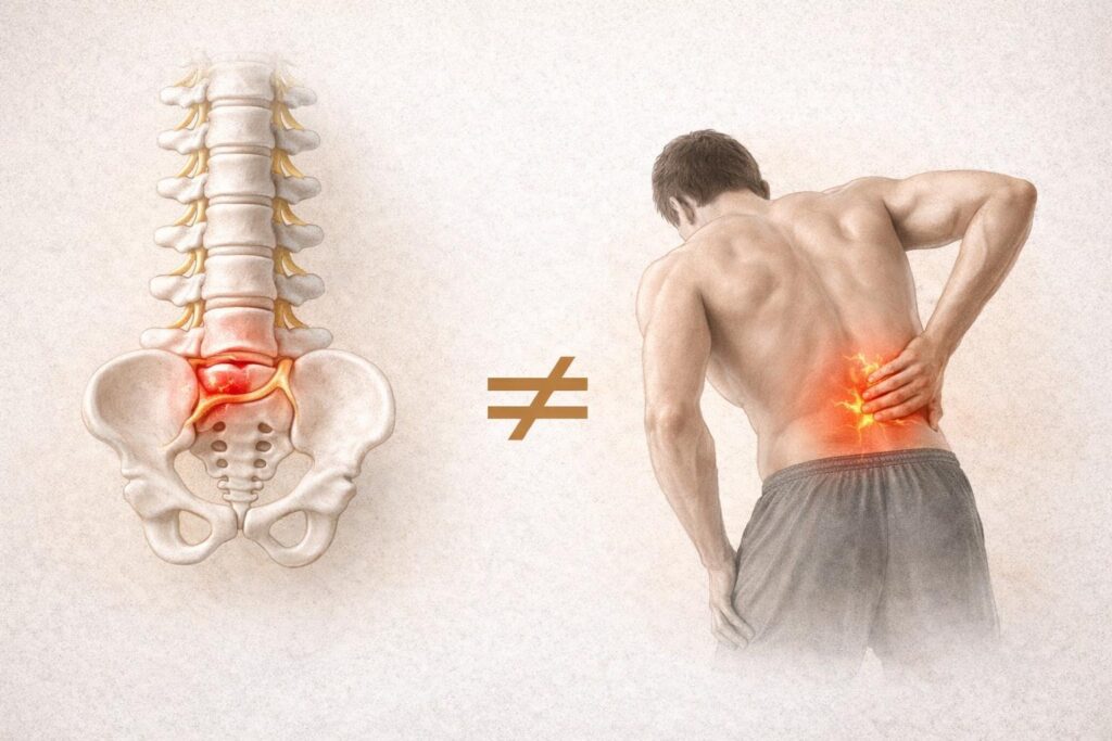
In modern healthcare systems, the first question that comes to mind for an individual consulting a specialist with low back pain is often: “Do I have a herniated disc?” The most commonly used method to answer this question is Magnetic Resonance Imaging (MRI). While seeing statements such as “disc herniation at the L4-L5 level” or “contact with the nerve root” in an MRI report may bring relief by seemingly identifying the source of pain, it often also introduces significant anxiety with the thought that “my back is damaged.” However, scientific literature points to a surprising and reassuring truth: structural changes identified in MRI scans may not always be the direct cause of pain.[1, 2, 3]
The long-standing belief that “damage equals pain” is being replaced by a much more dynamic and complex model. Large-scale studies have shown that thousands of people with no pain at all have herniations, osteoarthritis, and disc degeneration in their spines—just like individuals who experience low back pain.[4, 5, 6] This demonstrates that viewing low back pain purely as a mechanical failure is medically insufficient. The spine is not a static and fragile structure; it is a living system that continuously adapts, changes, and heals under the interaction of biological, psychological, and social factors.[7, 8] This report examines in detail the biological nature of disc herniation, the central role of the nervous system in pain perception, and how approaches to low back pain have evolved in light of scientific evidence.
Summary
A herniated disc is no longer considered an “inevitable cause of disability” in modern medicine. Scientific evidence has repeatedly demonstrated how resilient, adaptable, and capable of recovery the spine truly is.[5, 23, 31] MRI images provide us only with an anatomical “landscape”; however, what determines how painful that landscape will be is not just the size of the herniation, but also the sensitivity of our nervous system, our beliefs, and our overall health habits.[7, 8, 17]
For an individual, the key to managing low back pain is not to get lost in the technical terms of an MRI report, but to trust the body’s natural healing process and support it by staying active.[22, 29, 52] Herniations can shrink, nerves can calm down, and the brain can reset its pain threshold.[31, 32, 38] In summary, a herniated disc is not always the sole cause of pain, and in most cases, accurate knowledge and controlled movement are far more powerful healers than a surgeon’s scalpel.[25, 27, 29, 52]
The Anatomical Nature of Disc Herniation and Its Mechanisms
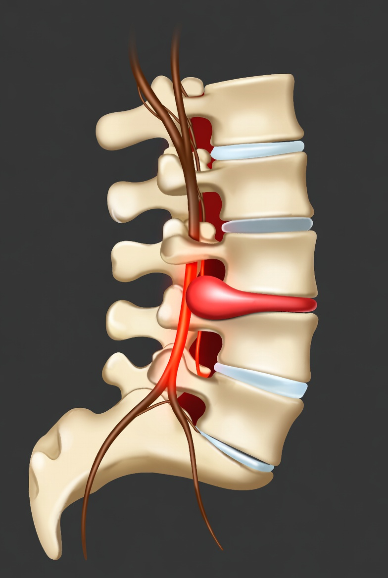
The spinal system is a complex structure that transfers the weight of the head through the pelvis to the legs and protects the spinal cord. The discs (intervertebral discs) located between the vertebrae are critical components that both enable mobility and absorb mechanical loads. The anatomy of a disc consists of two main parts designed to optimize mechanical forces: the gel-like nucleus pulposus at the center and the surrounding fibrous structure known as the annulus fibrosus.[9, 10]
A herniated disc, or lumbar disc herniation, is defined as the displacement of the nucleus pulposus material outward due to a weakness or tear in the surrounding annulus fibrosus fibers.[1, 11, 12] This process usually results not from a single acute injury but from a combination of years of microtrauma, genetic predisposition, and age-related changes in tissue quality.[9] Biomechanical studies show that combined forces of axial compression (vertical load), torsion (rotation), and flexion (forward bending) play a role in the development of disc herniation.[9]
Types of Disc Herniation and Clinical Definitions
| Clinical Definition | Radiological Correspondence | Pathological Process |
|---|---|---|
| Disk Bulging | General expansion of the disc without a complete tear in the fibrous structure | Loss of elasticity in fibers and spreading of the disc |
| Protrusion | The nucleus material gathers toward a focal point, but the outer fibers remain intact | Formation of a focal bulge, also known as the “early stage” of herniation |
| Extrusion | The nucleus material ruptures the fibrous ring and extends into the spinal canal | Stage where the immune system begins to respond |
| Sequestration | The fragment within the canal completely separates from the main disc structure | Although it appears most severe, it has the highest likelihood of natural resorption |
When these structural changes occur, the body does not perceive them merely as mechanical displacement. Disc material is recognized as a “foreign substance” by the immune system. Especially in extrusion and sequestration stages, immune cells (macrophages) migrate to the area and initiate an inflammatory process.[10, 13, 14] This inflammation can chemically irritate nerve roots and lead to pain.[9, 15] Therefore, disc-related pain is not only about “compression” of the nerve but also about exposure of the nerve to a “chemical environment.”
The Weak Relationship Between Imaging Findings and Pain
With the advancement of diagnostic methods in medicine, obtaining an MRI for almost every patient with low back pain has become a routine practice. However, the assumption that a herniation detected radiologically is the sole source of the patient’s pain is being seriously questioned by modern scientific evidence.[1, 2] MRI images provide a static snapshot of the spine at a given moment; however, pain is a dynamic process involving the nervous system, biochemical balance, and perceptual centers in the brain.[16, 17]
Systematic reviews in the literature have repeatedly demonstrated that there is only a moderate correlation between MRI findings and clinical symptoms.[1, 2, 3] For example, the presence of a “very large herniation” on an MRI report does not necessarily mean that the patient will experience severe pain. Similarly, individuals with completely “normal” MRI findings may suffer from disabling levels of low back pain.[17] This highlights the necessity of correlating radiological findings with the clinical picture; otherwise, misdiagnosis and unnecessary surgical interventions become inevitable.[3]
In one study, no statistically significant relationship was found between the degree of disc herniation, the level of degeneration, and functional disability scores (such as ODI and VAS).[2] In other words, how limited an individual is in daily life is more closely related to other factors (muscle strength, stress level, response to pain) than to the size of the herniation.[2, 18]
Disc Findings in Asymptomatic Individuals: “Wrinkles on the Inside”
One of the strongest pieces of scientific evidence regarding disc herniation comes from MRI studies conducted on healthy individuals with no low back pain. The large meta-analysis conducted by Brinjikji et al. (2015), involving 3,110 asymptomatic individuals, fundamentally changed our perspective on spinal findings.[4, 5, 19]
According to the data from this study, 37% of completely healthy individuals in their 20s have disc degeneration, and 30% have disc bulging.[5, 6] These rates increase dramatically with age. Among pain-free individuals aged 50, 4 out of 5 (80%) have disc degeneration.[5, 6, 20]
Table: Prevalence of Disc Findings by Age
| Age | Disc Degeneration (%) | Disc Bulging (%) | Disc Protrusion (%) |
|---|---|---|---|
| 20 | 37 | 30 | 29 |
| 30 | 52 | 40 | 31 |
| 40 | 68 | 50 | 33 |
| 50 | 80 | 60 | 36 |
| 60 | 88 | 69 | 38 |
| 70 | 93 | 77 | 40 |
| 80 | 96 | 84 | 43 |
The most important conclusion from these data is this: most of the changes occurring in the discs of your spine are not a “disease,” but rather a normal and expected part of the aging process.[5, 21] The most commonly used metaphor in the scientific community to explain this phenomenon is the “wrinkles on the inside” metaphor.[21, 22, 23]
Just as our skin wrinkles or our hair turns gray as we age, it is completely natural for spinal discs to lose fluid, bulge, or tear. Just as a wrinkle on the skin does not cause pain, these “normal” changes in the spine do not necessarily have to be a source of pain.[21, 22] For an individual encountering such findings, the most accurate medical diagnosis is often simply “being human” and “aging.”[22, 23]
The Reality That Low Back Pain Cannot Be Explained by a Structural Problem Alone
The traditional biomedical model, which dominated for decades, viewed the body as a machine and its organs as replaceable parts. According to this model, if something hurts, there must be a “fault” in that specific part. However, when it comes to low back pain, this model falls short.[7, 8] Low back pain is a multidimensional experience that cannot be explained solely by a disc fragment contacting a nerve on an MRI scan.[18]
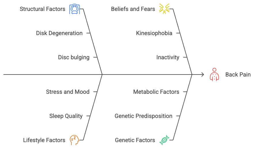
In many cases, pain persists even after the herniated disc fragment is surgically removed (Failed Back Surgery Syndrome), or conversely, pain completely resolves while the herniation remains in place. This proves that the source of pain is not solely “structural damage.”[1, 3, 24] Research shows that lifestyle factors play a more significant role in the onset and chronicity of low back pain than tissue damage itself.[7, 17]
Sleep Quality: Poor sleep lowers the body’s pain threshold and makes the nervous system more sensitive.[7, 17]
Stress and Mood: Chronic stress, anxiety, and depression disrupt pain-processing mechanisms in the brain, making even mild discomfort feel unbearable.[7, 8, 18]
Beliefs and Fears: The belief that “movement will cause more damage” (kinesiophobia) leads to inactivity, which results in muscle weakness and increased pain.[7, 25]
The metaphor of an overflowing cup effectively explains this phenomenon. The amount of water in the cup is not determined solely by the herniation (structural factor), but also by work stress, poor sleep, genetic factors, and lack of physical activity. When the cup overflows (i.e., when the body’s adaptive capacity is exceeded), the final drop (such as a simple bending movement) triggers pain. However, the real issue is not that “last drop,” but all the factors that filled the cup.[22]
The Explanatory Power of the Nervous System and the Biopsychosocial Model
Today, the approach to low back pain is built on the Biopsychosocial Model. This model defines pain as a complex interaction of biological tissue condition, psychological beliefs, and social environment.[7, 18] According to this model, pain is not a measure of tissue damage, but a “safety warning” produced by the brain to protect the body.[7, 26, 27]
Central Sensitization: The Nervous System in a High-Alert State
In individuals with chronic low back pain, pain may persist even after the tissues in the lower back have fully healed. One of the main reasons for this is central sensitization.[16, 17, 28] Central sensitization refers to a state in which the nervous system enters a “high alert” mode and begins to interpret normally non-painful stimuli (such as light touch or normal bending) as pain.[16, 26]
This phenomenon can be compared to a malfunctioning car alarm. Normally, the alarm should sound when a thief breaks a window. However, it becomes so sensitive that even a passing cat or a gust of wind triggers an ear-piercing alarm.[21, 26, 27] In such a patient, the primary target of treatment is not the lower back (the car window), but the alarm system itself (the nervous system).[16, 21]
Type of Pain Mechanism Clinical Feature Nociceptive Pain Actual or potential tissue damage Usually localized, movement-related Neuropathic Pain Disease or lesion in the nervous system Burning, electric shock, numbness Nociplastic Pain Altered pain processing in the nervous system Widespread pain, fatigue, sensitivity to touch (allodynia)
In cases of disc herniation, pain is usually a combination of these three types. Initially, tissue irritation caused by the herniation (nociceptive) and nerve root involvement (neuropathic) are dominant, but as the process continues, changes in the nervous system (nociplastic) become more prominent.[12, 26]
Scientific Evaluation of the Belief “If There Is a Herniation, There Must Be Pain”
There is a strong belief in society that “If I have a herniated disc, it is normal to have pain” or “My pain will not go away unless the herniation is fixed.” However, scientific studies clearly show that there is no linear relationship between the presence of a herniation and pain.[1, 2, 29]
Many individuals are able to return to a completely pain-free life through physiotherapy, exercise, and lifestyle changes, even though the herniated disc remains in place.[22, 24, 29] In some cases, follow-up MRIs taken after the pain has resolved still show the presence of the herniation. This indicates that the body has learned to live with the herniation, the nervous system has reduced its sensitivity in that area, and surrounding muscles have begun to better support the spine.[21, 29, 30]
On the other hand, surgically removing the herniation is not always a definitive solution. Surgery offers only a “mechanical” fix; however, if psychosocial factors, nervous system sensitivity, and faulty movement patterns that contribute to pain persist, the risk of recurrence after surgery is quite high.[7, 8] Scientific evidence shows that unless there are severe neurological deficits (such as loss of bladder control or progressive muscle weakness), conservative (non-surgical) treatments yield outcomes comparable to surgery in the long term.[24, 31]
The Self-Healing Potential of Herniated Discs: Spontaneous Regression
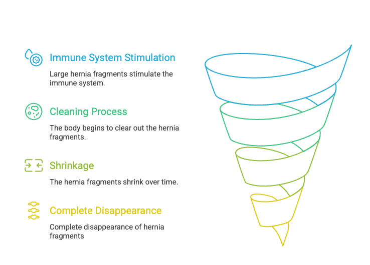
One of the greatest fears of patients diagnosed with a herniated disc is the belief that the herniation will remain permanently and can only be removed through surgery. However, the human body has an incredible self-repair mechanism. This phenomenon, known in the scientific literature as spontaneous regression, refers to the shrinking or complete disappearance of the herniated disc material over time.[31, 32, 33]
Interestingly, the larger the herniation (the more it extends into the spinal canal), the higher the likelihood that it will be recognized and resorbed by the body.[13, 32, 33] This is because larger fragments act as a stronger stimulus for the immune system, triggering a more active “clean-up” process.[10, 13, 31]
Table: Spontaneous Regression Rates of Herniated Discs
| Herniation Type | Spontaneous Regression Rate | Complete Resolution Rate |
|---|---|---|
| Sequestration (Free fragment) | 96% | 43% |
| Extrusion | 70% | 15% |
| Protrusion | 41% | Insufficient data |
| Bulging | 13% | Insufficient data |
The systematic review by Chiu et al. (2015) demonstrated that free disc fragments regress spontaneously at an extraordinary rate of 96%.[32, 33] This process of shrinkage typically begins within 6 to 12 weeks and occurs through phagocytosis (the body “digesting” the fragment) and dehydration (loss of fluid leading to shrinkage).[13, 14, 34, 35] These findings highlight the importance of allowing the body time to heal rather than rushing into surgery.[31, 35]
Clinical Implications: A New Era in Physiotherapy and Patient Management
These scientific realities are fundamentally changing how patients with low back pain are managed. The focus is no longer on “eliminating the herniation,” but on healing and strengthening the individual.[7, 8, 25]
The Nocebo Effect of Diagnostic Reports and Communication
The technical language used in imaging reports (degeneration, tear, collapse) can create the perception in patients that “my back is ruined,” leading to a nocebo effect.[29, 36, 37] The nocebo effect refers to a situation where negative expectations increase a patient’s pain and level of disability.[29, 36]
When informing a patient about a herniation, explaining that it is often a normal part of aging and that it can heal spontaneously is one of the most important factors that accelerates recovery.[25, 29, 38] The language used by radiologists and clinicians directly affects a patient’s willingness to move. Studies show that reducing fear-inducing language in reports decreases unnecessary invasive procedures and lowers anxiety levels.[36, 37, 39]
The Importance of Movement and Physical Activity
The previously recommended “bed rest” for low back pain is now considered a medical mistake.[40, 41, 42] Cochrane reviews have shown that bed rest does not accelerate recovery; on the contrary, it weakens muscles, slows circulation, and prolongs recovery time.[40, 43]
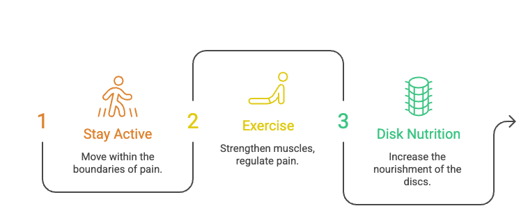
The modern approach recommends staying active within pain limits and gradually loading the spine.[40, 44, 45] Exercise not only strengthens muscles but also regulates pain centers in the brain and promotes the release of natural pain-relieving hormones (endorphins).[21, 25] Activities such as walking, swimming, yoga, or controlled strength training support the healing process by improving disc nutrition (via diffusion).[21, 45]
Pain Neuroscience Education (PNE)
One of the most important components of physiotherapy is helping patients understand how pain works.[25, 38] Pain Neuroscience Education (PNE) explains that pain does not always indicate tissue damage, why the nervous system can become sensitized, and how stress can increase pain.[25, 46]
Through this education, patients begin to fear their pain less, which reduces kinesiophobia (fear of movement) and leads to a more active and successful rehabilitation process.[25, 38]
When Is Surgery Necessary? “Red Flags”
Although more than 90% of low back pain cases recover without surgery, approximately 1–2% may involve serious medical conditions.[7, 47, 48] These are identified using clinical warning signs known as “Red Flags.” In the presence of these signs, immediate medical attention is required.[47, 49, 50]
Table: Red Flags
| Symptom / Condition | Urgency and Cause |
|---|---|
| Loss of bladder or bowel control (incontinence or retention) | Cauda Equina Syndrome – Requires emergency surgery |
| Numbness in the perineal (saddle) region | Indicates severe nerve root compression |
| Sudden and progressive muscle weakness in the legs | Risk of permanent nerve damage |
| Severe pain that wakes from sleep and does not improve with rest | Suggests malignancy (tumor) or infection |
| Unexplained weight loss and high fever | Indicates systemic disease or infection |
In the absence of these rare and serious signs, the first-line treatment for low back pain or radiating leg pain (sciatica) should always be non-surgical approaches.[31, 47, 51]
References
- Clinico Radiological Correlation of Radiculopathy in Lower Back Ache Patient With MRI, https://healthcare-bulletin.co.uk/article/clinico-radiological-correlation-of-radiculopathy-in-lower-back-ache-patient-with-mri-2769/
- Clinical correlation of magnetic resonance imaging with symptom complex in prolapsed intervertebral disc disease: A cross-sectional double blind analysis – PMC, https://pmc.ncbi.nlm.nih.gov/articles/PMC3872655/
- Correlation between clinical features and magnetic resonance imaging findings in lumbar disc prolapse – PMC, https://pmc.ncbi.nlm.nih.gov/articles/PMC2911925/
- Systematic Literature Review of Imaging Features of Spinal Degeneration in Asymptomatic Populations – Semantic Scholar, https://www.semanticscholar.org/paper/Systematic-Literature-Review-of-Imaging-Features-of-Brinjikji-Luetmer/84c5cda9173813a45c3a48adb8154ef25ac644cb
- Systematic Literature Review of Imaging Features of Spinal …, https://pmc.ncbi.nlm.nih.gov/articles/PMC4464797/
- Systematic Literature Review of Imaging Features of Spinal Degeneration in Asymptomatic Populations | American Journal of Neuroradiology, https://www.ajnr.org/content/36/4/811.short
- Acute low back pain, http://pain-ed.com/wp-content/uploads/2014/02/Osullivan-and-Lin-Pain-management-today-2014.pdf
- Unraveling the Complexity of Low Back Pain | Journal of Orthopaedic & Sports Physical Therapy – jospt, https://www.jospt.org/doi/10.2519/jospt.2016.0609
- The Biomechanical Landscape of Lumbar Disc Herniation: Mechanobiological Insights Into Injury and Regeneration – PMC, https://pmc.ncbi.nlm.nih.gov/articles/PMC12890390/
- Biological Mechanisms of Pain Management in Lumbar Disk Herniation: Focus on Cytokine Correlations and Therapeutic Approaches – ResearchGate, https://www.researchgate.net/publication/397393501_Biological_Mechanisms_of_Pain_Management_in_Lumbar_Disk_Herniation_Focus_on_Cytokine_Correlations_and_Therapeutic_Approaches
- Association Between Lumbar Disc Herniation and Vertebral Endplate Modic Changes: An MRI-Based Study – PMC, https://pmc.ncbi.nlm.nih.gov/articles/PMC12543248/
- Biological Mechanisms of Pain Management in Lumbar Disk Herniation: Focus on Cytokine Correlations and Therapeutic Approaches – PMC, https://pmc.ncbi.nlm.nih.gov/articles/PMC12652388/
- The probability of spontaneous regression of lumbar herniated disc: a systematic review, https://www.researchgate.net/publication/263815470_The_probability_of_spontaneous_regression_of_lumbar_herniated_disc_a_systematic_review
- Bel Fıtığı Kendi Kendine Geçer Mi? Tedavi Yöntemleri ve Öneriler – Dr. Mehmet Sorar, https://www.drsorar.com/blog-detay/belfitigi-kendi-kendine-gecer-mi
- Chemical and mechanical nerve root insults induce differential behavioral sensitivity and glial activation that are enhanced in combination – PMC, https://pmc.ncbi.nlm.nih.gov/articles/PMC2174426/
- CENTRAL SENSITIZATION IN CHRONIC PAIN: NEUROPHYSIOLOGICAL MECHANISMS AND CLINICAL IMPLICATIONS | International Journal of Innovative Technologies in Social Science, https://rsglobal.pl/index.php/ijitss/article/view/4428
- Is the Central Sensitization in Chronic Nonspecific Low Back Pain Structural Phenomenon or Psychological Reaction? A Narrative Review – MDPI, https://www.mdpi.com/2077-0383/14/2/577
- Chronic Low Back Pain: Bio-Psycho-Social Model – Core Concepts, https://www.coreconceptsphysio.sg/article/chronic-low-back-pain-bio-psycho-social-model-in-physiotherapy-treatment/
- (Open Access) Systematic Literature Review of Imaging Features of Spinal Degeneration in Asymptomatic Populations (2015) | Waleed Brinjikji | 900 Citations – SciSpace, https://scispace.com/papers/systematic-literature-review-of-imaging-features-of-spinal-38u81928sn?citations_page=153
- Systematic literature review of imaging features of spinal degeneration in asymptomatic populations – PubMed, https://pubmed.ncbi.nlm.nih.gov/25430861/
- Hurt vs. Harm: Understanding Pain – Physical Therapy, https://healingmotionpt.com/2021/02/20/hurt-vs-harm-understanding-pain/
- Tissue changes and pain: explaining their relevance – Greg Lehman, https://www.greglehman.ca/blog/2017/3/6/tissue-changes-and-pain-explaining-their-relevance
- Barbell Medicine Podcast | RedCircle, https://redcircle.com/shows/barbell-medicine-podcast/ep/fadf6bcf-21bb-42b1-882f-4f5bf3c80975
- Spontaneous resorption of herniated lumbar discs: illustrative cases – PMC, https://pmc.ncbi.nlm.nih.gov/articles/PMC12278955/
- Pain Neuroscience Education for Acute Pain – International Journal of Sports Physical Therapy, https://ijspt.org/wp-content/uploads/2024/05/10-Louw.pdf
- Central Sensitization: When It Is Not “All in Your Head” | AAFP, https://www.aafp.org/pubs/afp/issues/2023/0100/curbside-central-sensitization.html
- back pain — Blog — Kinfolk Physiotherapy & Wellness, https://www.kinfolkwellness.com.au/blog/tag/back+pain
- Central Sensitization in Chronic Low Back Pain: A Population-Based Study of a Japanese Mountain Village – PubMed, https://pubmed.ncbi.nlm.nih.gov/34040431/
- The Nocebo Effect: How Your MRI Report Can Hurt You – One on One Physical Therapy Atlanta GA, https://onetherapy.com/the-nocebo-effect/
- Signs a Herniated Disc is Healing: Timeline & Recovery – Spine MD, https://www.spine.md/insights/signs-herniated-disc-is-healing
- Spontaneous Resorption of Lumbar Disc Herniation: A Narrative Review of Pathophysiology, Predictive Factors, and Clinical Decision-Making – MDPI, https://www.mdpi.com/2673-4087/7/2/30
- The probability of spontaneous regression of lumbar herniated disc: a systematic review, https://pubmed.ncbi.nlm.nih.gov/25009200/
- The Probability of Spontaneous Regression of Lumbar Herniated Disc – A Systematic Review – Scribd, https://www.scribd.com/document/617906662/The-probability-of-spontaneous-regression-of-lumbar-herniated-disc-a-systematic-review
- Spontaneous regression of lumbar disc herniation: four cases report and review of the literature – PMC, https://pmc.ncbi.nlm.nih.gov/articles/PMC11439616/
- Herniated disks unchanged over time: Size reduced after oxygen–ozone therapy – PMC, https://pmc.ncbi.nlm.nih.gov/articles/PMC4984384/
- MRI Reporting for Lower Back Pain · Info for Participants · Clinical Trial 2026 | Power, https://www.withpower.com/trial/phase-nocebo-effects-11-2023-f22eb
- Study Details | NCT06103474 | Clinical Reporting to Alleviate the Nocebo Effect | ClinicalTrials.gov, https://clinicaltrials.gov/study/NCT06103474
- Effectiveness of Pain Neuroscience Education in Reducing Pain, Disability, Kinesiophobia, and Catastrophizing in Patients with Chronic Low Back Pain: A Systematic Review and Meta-Analysis – MDPI, https://www.mdpi.com/2076-3271/13/4/290
- Low Back Pain Patients’ Perceptions Regarding Their Own Radiology Reports – PMC – NIH, https://pmc.ncbi.nlm.nih.gov/articles/PMC10029370/
- The updated cochrane review of bed rest for low back pain and sciatica – PubMed, https://pubmed.ncbi.nlm.nih.gov/15738787/
- The Cochrane review of bed rest for acute low back pain and sciatica – PubMed, https://pubmed.ncbi.nlm.nih.gov/11074682/
- Advice to rest in bed versus advice to stay active for acute low-back pain and sciatica, https://pubmed.ncbi.nlm.nih.gov/20556780/
- Advice to rest in bed versus advice to stay active for acute low‐back pain and sciatica – PMC, https://pmc.ncbi.nlm.nih.gov/articles/PMC12161166/
- For Acute Low Back Pain, Staying Active May Be Better Than Bed Rest – Medscape Education, https://www.medscape.org/viewarticle/724386
- Bel Fıtığı Belirtileri Nelerdir? Bel Fıtığı Nasıl Geçer? – Acıbadem, https://www.acibadem.com.tr/ilgi-alani/bel-fitigi/
- Know Pain, Know Gain? A Perspective on Pain Neuroscience Education in Physical Therapy – JOSPT.org, https://www.jospt.org/doi/10.2519/jospt.2016.0602
- Low back pain imaging – EBI – AOMRC, https://ebi.aomrc.org.uk/interventions/low-back-pain-imaging-2/
- Bel ağrısı tanı ve tedavisinde kırmızı ve mavi bayraklar – TOTBİD Derleme Dergisi, https://totbidderlemedergisi.org/pdf.php?id=834
- When Is Imaging Appropriate for a Patient With Low Back Pain? – PMC – NIH, https://pmc.ncbi.nlm.nih.gov/articles/PMC7529135/
- Revisiting the “Red Flags” in Acute Low Back Pain | Consultant360, https://www.consultant360.com/peer-reviewed/revisiting-red-flags-acute-low-back-pain
- Low Back Pain – Preview – American College of Radiology, https://acsearch.acr.org/docs/69483/narrative/
- Bel Fıtığı Geçer mi? Bilim Ne Diyor?- Kadıköy Fizik Tedavi, https://mterapi.com/bel-fitigi-gecer-mi-ameliyatsiz-tedavi/